Copyright
©2013 Baishideng Publishing Group Co.
World J Radiol. May 28, 2013; 5(5): 193-201
Published online May 28, 2013. doi: 10.4329/wjr.v5.i5.193
Published online May 28, 2013. doi: 10.4329/wjr.v5.i5.193
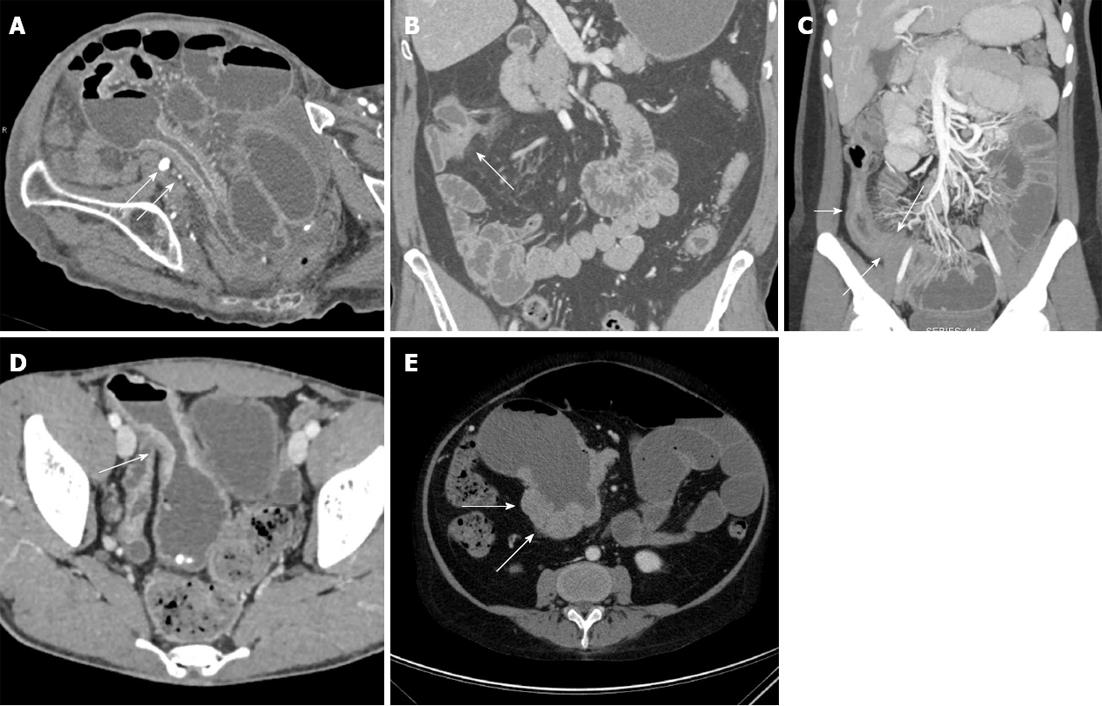
Figure 3 Bowel-related complications of Crohn’s disease.
A: Eighty-four-year-old who presented with abdominal pain. Axial contrast-enhanced image demonstrates multiple dilated loops of small bowel, in keeping with a small bowel obstruction. This image also demonstrates a long segment of bowel wall thickening and mucosal hyperemia with luminal narrowing (arrows). Given the patient’s age, this was originally thought to represent a neoplastic or ischemic stricture, but was found to represent late-onset Crohn’s disease after surgery; B: Forty-nine-year-old male with Crohn’s disease. Coronal volume rendered computed tomography image demonstrates a short segment stricture and focal thickening of the hepatic flexure of the colon, with minimal adjacent induration, but no significant mesenteric hyperemia. This was thought to be a chronic-appearing stricture. Colonoscopy was performed to exclude an underlying neoplasm, and the patient was found to have a chronic stricture in this location without acute inflammation or evidence of tumor; C: Twenty-four-year-old female with Crohn’s disease. Coronal volume rendered image demonstrates several dilated loops of small bowel in the left abdomen, with a discrete transition point (long arrows) in the distal ileum, at the site of a long segment of narrowed, hyperemic, thickened, inflamed small bowel (short arrow); D: Twenty-five-year-old male with Crohn’s disease. Axial contrast enhanced image demonstrates thickening of a loop of small bowel in the pelvis, with dilatation proximal to the site of stricturing. Given the irregular thickening (arrow) at this site, the possibility of a malignancy could not be excluded. As a result, the patient underwent surgical resection and was found to have a small bowel adenocarcinoma; E: Fifty-one-year-old female with a history of Crohn’s disease. Axial image demonstrates nodular soft tissue thickening (arrows) surrounding an aneurysmally dilated loop of bowel in the right abdomen. This was found to represent B-cell lymphoma following surgical resection.
- Citation: Raman SP, Horton KM, Fishman EK. Computed tomography of Crohn’s disease: The role of three dimensional technique. World J Radiol 2013; 5(5): 193-201
- URL: https://www.wjgnet.com/1949-8470/full/v5/i5/193.htm
- DOI: https://dx.doi.org/10.4329/wjr.v5.i5.193


