Copyright
©2013 Baishideng Publishing Group Co.
World J Radiol. May 28, 2013; 5(5): 193-201
Published online May 28, 2013. doi: 10.4329/wjr.v5.i5.193
Published online May 28, 2013. doi: 10.4329/wjr.v5.i5.193
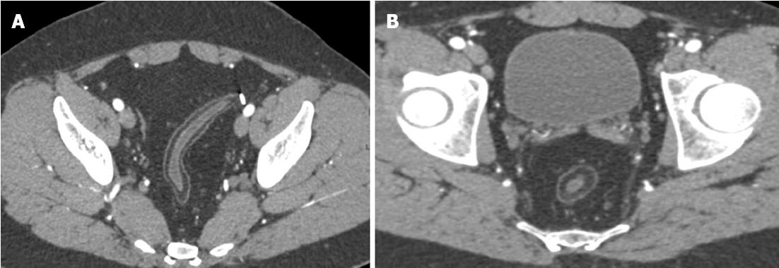
Figure 2 Sequela of chronic Crohn’s related bowel inflammation.
Twenty-seven year-old male with Crohn’s disease. Axial images demonstrate diffuse fat deposition in the wall of the rectosigmoid colon (A, B), as well as marked fibrofatty proliferation (“creeping fat”) (B) surrounding the rectum.
- Citation: Raman SP, Horton KM, Fishman EK. Computed tomography of Crohn’s disease: The role of three dimensional technique. World J Radiol 2013; 5(5): 193-201
- URL: https://www.wjgnet.com/1949-8470/full/v5/i5/193.htm
- DOI: https://dx.doi.org/10.4329/wjr.v5.i5.193


