Copyright
©2013 Baishideng Publishing Group Co.
World J Radiol. May 28, 2013; 5(5): 193-201
Published online May 28, 2013. doi: 10.4329/wjr.v5.i5.193
Published online May 28, 2013. doi: 10.4329/wjr.v5.i5.193
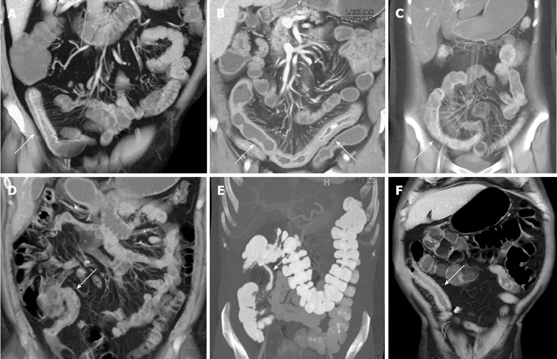
Figure 1 Active Crohn’s disease.
A: Fifty-four-year-old male with Crohn’s disease. Coronal volume rendered image demonstrates prominent wall thickening and mucosal hyperemia encompassing a 10 cm segment of ileum (arrow). The volume rendered three dimensional (3-D) image nicely accentuates the marked mesenteric hyperemia and vasa recta engorgement adjacent to the inflamed loop of bowel; B: Sixty-two-year-old male with Crohn’s disease. Coronal volume rendered images demonstrate a long segment of markedly thickened, inflamed bowel in the pelvis (arrows). Notably, the 3-D images accentuate the marked engorgement of the vasa recta (“comb sign”) adjacent to the inflamed loop of bowel; C: Twenty-two-year-old with Crohn’s disease. Coronal volume rendered image demonstrates an acutely inflamed loop of colon (arrow) with mucosal hyperemia and wall thickening, as well as adjacent engorgement of the vasa recta; D: Thirty-eight-year-old male with Crohn’s disease. Coronal volume rendered image demonstrates thickening and mucosal hyperemia of the terminal ileum, a classic appearance and location for acute Crohn’s related inflammation; E: Thirty-four-year-old male with Crohn’s disease. Coronal volume rendered image demonstrates thickening of an intermediate length segment of terminal ileum (arrow); F: Sixty-year-old male with Crohn’s disease and history of prior ileal resection and reanastomosis. Coronal volume rendered image demonstrates marked thickening, mucosal hyperemia, and adjacent vasa recta engorgement of the neo-terminal ileum (arrow).
- Citation: Raman SP, Horton KM, Fishman EK. Computed tomography of Crohn’s disease: The role of three dimensional technique. World J Radiol 2013; 5(5): 193-201
- URL: https://www.wjgnet.com/1949-8470/full/v5/i5/193.htm
- DOI: https://dx.doi.org/10.4329/wjr.v5.i5.193


