Copyright
©2013 Baishideng Publishing Group Co.
World J Radiol. Apr 28, 2013; 5(4): 156-165
Published online Apr 28, 2013. doi: 10.4329/wjr.v5.i4.156
Published online Apr 28, 2013. doi: 10.4329/wjr.v5.i4.156
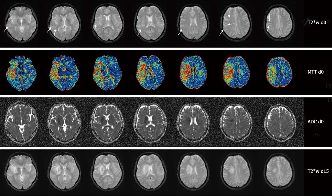
Figure 2 Image montage of magnetic resonance imaging findings in a 46-year-old woman with a right middle cerebral artery occlusion 321 min after symptom onset and an National Institute of Health Stroke Score of 11 on admission.
Note the subtle asymmetrically prominent veins (arrows) on T2*w imaging (first row) within the mean transit time (MTT) lesion (second row) but outside the apparent diffusion coefficient (ADC) lesion (third row) on the day of admission (d0). On follow-up imaging 15 d later (d15) vessel appearance has normalized (fourth row).
- Citation: Jensen-Kondering U, Böhm R. Asymmetrically hypointense veins on T2*w imaging and susceptibility-weighted imaging in ischemic stroke. World J Radiol 2013; 5(4): 156-165
- URL: https://www.wjgnet.com/1949-8470/full/v5/i4/156.htm
- DOI: https://dx.doi.org/10.4329/wjr.v5.i4.156


