Copyright
©2013 Baishideng Publishing Group Co.
World J Radiol. Apr 28, 2013; 5(4): 143-155
Published online Apr 28, 2013. doi: 10.4329/wjr.v5.i4.143
Published online Apr 28, 2013. doi: 10.4329/wjr.v5.i4.143
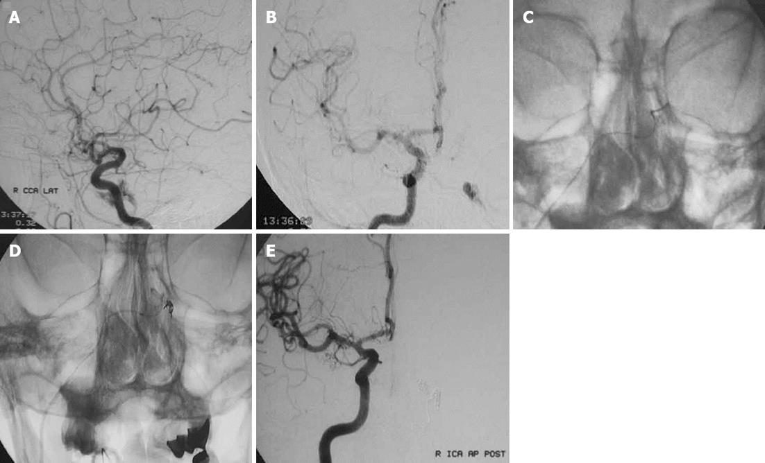
Figure 4 A forty-year-old woman with chemosis of the left eye and diplopia was found to have a dural carotid cavernous sinus fistula.
A, B: Frontal (A) and lateral (B) injections of the right common carotid artery demonstrating left dural carotid cavernous sinus fistula with antegrade drainage; C: Access to the fistula site through the contralateral (right inferior petrous sinus) transvenous route and positioning of the microcatheter; D: Coil embolization within the microcather extending into the venous compartment of the fistula; E: Posttreatment frontal digital subtraction angiogram view of right internal carotid artery demonstrating obliteration of the fistula and lack of residual filling.
- Citation: Korkmazer B, Kocak B, Tureci E, Islak C, Kocer N, Kizilkilic O. Endovascular treatment of carotid cavernous sinus fistula: A systematic review. World J Radiol 2013; 5(4): 143-155
- URL: https://www.wjgnet.com/1949-8470/full/v5/i4/143.htm
- DOI: https://dx.doi.org/10.4329/wjr.v5.i4.143


