Copyright
©2013 Baishideng Publishing Group Co.
World J Radiol. Apr 28, 2013; 5(4): 143-155
Published online Apr 28, 2013. doi: 10.4329/wjr.v5.i4.143
Published online Apr 28, 2013. doi: 10.4329/wjr.v5.i4.143
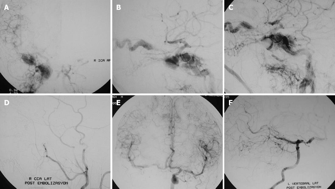
Figure 3 A thirty one-year-old male patient with right ophthalmoplegia following head trauma was found to have a direct carotid cavernous sinus fistula.
A, B: Frontal (A) and lateral (B) digital subtraction angiogram views of right internal carotid artery (ICA) demonstrating laceration of ICA, pseudoaneurysm in the cavernous ICA and direct carotid cavernous sinus fistula; C: After considering the presence of the pseudoaneurysm, two detachable balloons were positioned to occlude the parent artery; D: Right CCA digital subtraction angiogram after balloon occlusion of the ICA showing complete obliteration of the fistula; E, F: Posttreatment left ICA (E) and left vertebral artery (F) injections demonstrating reconstruction of the right ICA area.
- Citation: Korkmazer B, Kocak B, Tureci E, Islak C, Kocer N, Kizilkilic O. Endovascular treatment of carotid cavernous sinus fistula: A systematic review. World J Radiol 2013; 5(4): 143-155
- URL: https://www.wjgnet.com/1949-8470/full/v5/i4/143.htm
- DOI: https://dx.doi.org/10.4329/wjr.v5.i4.143


