Copyright
©2013 Baishideng Publishing Group Co.
World J Radiol. Apr 28, 2013; 5(4): 143-155
Published online Apr 28, 2013. doi: 10.4329/wjr.v5.i4.143
Published online Apr 28, 2013. doi: 10.4329/wjr.v5.i4.143
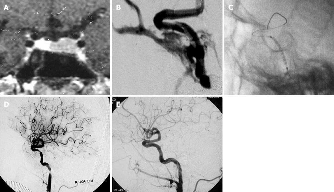
Figure 2 A fifty two-year-old woman who presented with galactorrhea due to hypophyseal adenoma underwent transsphenoidal surgery.
During the operation internal carotid artery laceration and massive arterial hemorrhage occurred. A: T1 W C+ coronal magnetic resonance imaging view demonstrating hypointensity at the left hypophyseal region which was consistent with hypophyseal adenoma; B: Immediate defect source analysis revealed a defect at the anteromedial wall of right internal carotid artery (ICA) and associated carotid cavernous sinus fistula; C: Position of the stent-graft closing the orifice of the fistula; D: Postprocedural right ICA injection demonstrating complete obliteration of the fistula and concentric luminal stenosis at the petrous segment associated with mechanic vasospasm secondary to guide-wire and stent manipulation; E: Third month control angiography revealed regular parent artery contours, absence of recurrent fistula filling and intimal hyperplasia within the stent.
- Citation: Korkmazer B, Kocak B, Tureci E, Islak C, Kocer N, Kizilkilic O. Endovascular treatment of carotid cavernous sinus fistula: A systematic review. World J Radiol 2013; 5(4): 143-155
- URL: https://www.wjgnet.com/1949-8470/full/v5/i4/143.htm
- DOI: https://dx.doi.org/10.4329/wjr.v5.i4.143


