Copyright
©2013 Baishideng.
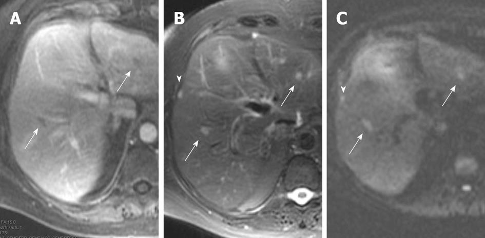
Figure 7 Magnetic resonance imaging images from patient with liver metastases from biopsy proven pancreatic cancer.
A: Portal venous phase image, shows two hypointense foci (arrows) within the liver; B: T2 fat suppressed images also show the two lesions and show to better effect the subcapsular metastasis (arrowhead), arrows are metastases in liver parenchyma; C: Diffusion weighted image (b value of 500 s/mm2) shows all three lesions well, without confounding T2 bright signal in biliary tree or vessels seen on T2 weighted images. Arrows are metastases in liver parenchyma, arrowhead is the subcapsular liver metastasis.
- Citation: Tamm EP, Bhosale PR, Vikram R, de Almeida Marcal LP, Balachandran A. Imaging of pancreatic ductal adenocarcinoma: State of the art. World J Radiol 2013; 5(3): 98-105
- URL: https://www.wjgnet.com/1949-8470/full/v5/i3/98.htm
- DOI: https://dx.doi.org/10.4329/wjr.v5.i3.98


