Copyright
©2013 Baishideng.
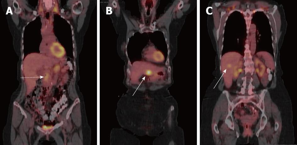
Figure 5 Coronal fused positron emission tomography with computed tomography images of patient with pancreatic cancer and liver metastases.
A: Primary tumor shows mild-moderate uptake (dashed arrow); B, C: While liver metastases in (B) and (C) show variable uptake (solid arrows).
- Citation: Tamm EP, Bhosale PR, Vikram R, de Almeida Marcal LP, Balachandran A. Imaging of pancreatic ductal adenocarcinoma: State of the art. World J Radiol 2013; 5(3): 98-105
- URL: https://www.wjgnet.com/1949-8470/full/v5/i3/98.htm
- DOI: https://dx.doi.org/10.4329/wjr.v5.i3.98


