Copyright
©2013 Baishideng.
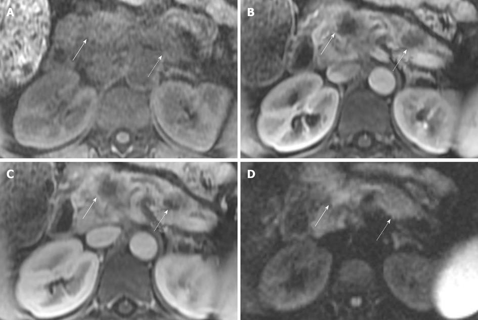
Figure 4 Magnetic resonance imaging of patient with two sites of pancreatic cancer.
A: T1 fat suppressed images do not show the tumor well in this patient, but are often helpful. Solid arrow indicates the pancreatic neck tumor, dashed arrow indicates the pancreatic tail tumor; B, C: However, both sites are well seen, in the neck (solid arrows) and tail (dashed arrows) on the pancreatic parenchymal phase (B) and the portal venous phase (C); D: In this case sites of tumor are not well seen on diffusion weighted imaging. Solid arrow indicates the pancreatic neck tumor, dashed arrow indicates the pancreatic tail tumor. Conspicuity of the primary lesion can be very variable on diffusion weighted imaging.
- Citation: Tamm EP, Bhosale PR, Vikram R, de Almeida Marcal LP, Balachandran A. Imaging of pancreatic ductal adenocarcinoma: State of the art. World J Radiol 2013; 5(3): 98-105
- URL: https://www.wjgnet.com/1949-8470/full/v5/i3/98.htm
- DOI: https://dx.doi.org/10.4329/wjr.v5.i3.98


