Copyright
©2013 Baishideng.
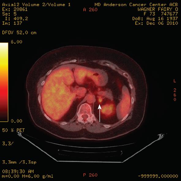
Figure 9 A positron emission tomography/computed tomography fused axial image demonstrates a left adrenal mass (arrow) with increased fluorodeoxyglucose activity slightly greater than the liver in a patient with non small cell lung cancer.
Biopsy revealed an adenoma, rendering this a false positive finding.
- Citation: Korivi BR, Elsayes KM. Cross-sectional imaging work-up of adrenal masses. World J Radiol 2013; 5(3): 88-97
- URL: https://www.wjgnet.com/1949-8470/full/v5/i3/88.htm
- DOI: https://dx.doi.org/10.4329/wjr.v5.i3.88


