Copyright
©2013 Baishideng.
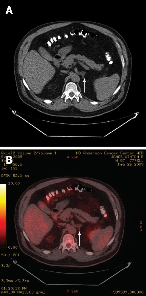
Figure 7 Axial non-contrast computed tomography image (A) and fused positron emission tomography/computed tomography image (B) of a patient with esophageal cancer with a left adrenal adenoma (arrows).
The adenoma has an Hounsfield Units of -10 on the non-contrast computed tomography (A) and demonstrates fluorodeoxyglucose uptake less than the liver (B), and is therefore not consistent with a metastasis.
- Citation: Korivi BR, Elsayes KM. Cross-sectional imaging work-up of adrenal masses. World J Radiol 2013; 5(3): 88-97
- URL: https://www.wjgnet.com/1949-8470/full/v5/i3/88.htm
- DOI: https://dx.doi.org/10.4329/wjr.v5.i3.88


