Copyright
©2013 Baishideng.
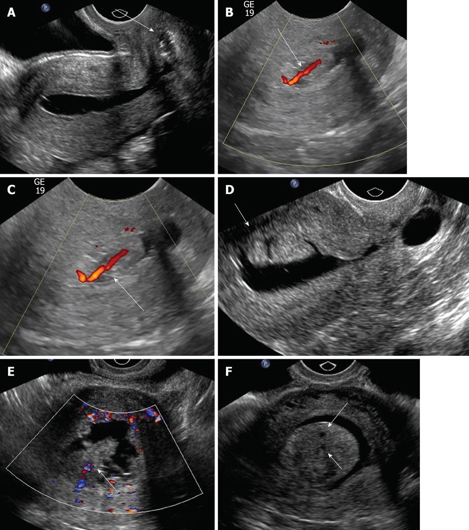
Figure 3 Multiple manifestations of endometrial polyps.
A: Sonohysterographical image of a cervical polyp (arrow); B, C: Sonohysterograms with Doppler demonstrating endometrial polyps with feeding vessels (arrows); D: Sonohysterogram shows elongated bilobed mass (arrow) attached to endometrial and projecting into the endometrial canal, representing an endometrial polyp; E: Broad based, hypoechoic, slightly hypervascular mass with slightly jagged borders projecting into the endometrial canal (arrow); F: Sonohysterogram shows single, echogenic polypoid lesion, representing an endometrial polyp arising with several tiny cysts (arrow).
- Citation: Yang T, Pandya A, Marcal L, Bude RO, Platt JF, Bedi DG, Elsayes KM. Sonohysterography: Principles, technique and role in diagnosis of endometrial pathology. World J Radiol 2013; 5(3): 81-87
- URL: https://www.wjgnet.com/1949-8470/full/v5/i3/81.htm
- DOI: https://dx.doi.org/10.4329/wjr.v5.i3.81


