Copyright
©2013 Baishideng.
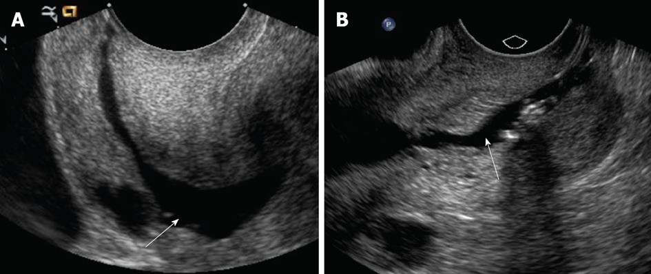
Figure 1 Procedural steps: Fluid instillation into the endometrial cavity (A, B).
Longitudinal ultrasound images of the lower uterine segment show adequate distention of the endocervical canal with warm saline during sonohysterography (arrows). During this phase, it is critical to observe for over or underdistention of the endocervical canal.
- Citation: Yang T, Pandya A, Marcal L, Bude RO, Platt JF, Bedi DG, Elsayes KM. Sonohysterography: Principles, technique and role in diagnosis of endometrial pathology. World J Radiol 2013; 5(3): 81-87
- URL: https://www.wjgnet.com/1949-8470/full/v5/i3/81.htm
- DOI: https://dx.doi.org/10.4329/wjr.v5.i3.81


