Copyright
©2013 Baishideng.
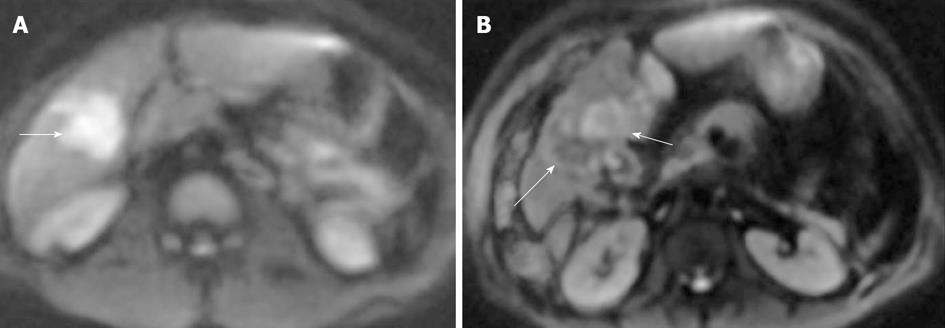
Figure 9 Hepatocellular carcinoma (arrows) on diffusion-weighted image (A) before and (B) after transarterial chemoembolization.
A: Hepatocellular carcinoma (HCC) was hyperintense compared with the cirrhotic liver on diffusion weighted image, with an apparent diffusion coefficient (ADC) = 1.41 × 10⁻³ mm2/s vs 1.08 × 10⁻³ mm2/s for the liver; B: After transarterial chemoembolization, HCC showed a residual area of hyperintense signal (short arrow) with ADC = 1.38 × 10⁻³ mm2/s (residual tumor) and a new area of hypointense signal (long arrow) with ADC = 1.87 × 10⁻³ mm2/s (coagulation necrosis), indicating a partial response. This finding of necrosis was supported by lack of enhancement on gadolinium-enhanced imaging and confirmed on follow-up studies.
- Citation: Morani AC, Elsayes KM, Liu PS, Weadock WJ, Szklaruk J, Dillman JR, Khan A, Chenevert TL, Hussain HK. Abdominal applications of diffusion-weighted magnetic resonance imaging: Where do we stand. World J Radiol 2013; 5(3): 68-80
- URL: https://www.wjgnet.com/1949-8470/full/v5/i3/68.htm
- DOI: https://dx.doi.org/10.4329/wjr.v5.i3.68


