Copyright
©2013 Baishideng.
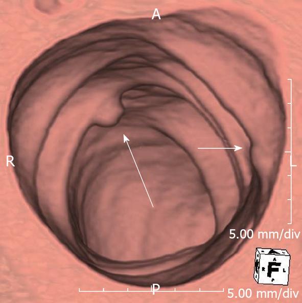
Figure 5 A 72-year-old male underwent computed tomography colonoscopy.
There is an 8 mm polyp in descending colon (long arrow), which is easily detected in the 3D view. Note that there is another ‘’polyp’’ like lesion (short arrow). This was shown to be a fecal material by correlating with the 2D view. False positives for polyp are one of the pitfalls of 3D review but this can be easily overcome by complementing 3D with 2D review.
- Citation: Ganeshan D, Elsayes KM, Vining D. Virtual colonoscopy: Utility, impact and overview. World J Radiol 2013; 5(3): 61-67
- URL: https://www.wjgnet.com/1949-8470/full/v5/i3/61.htm
- DOI: https://dx.doi.org/10.4329/wjr.v5.i3.61


