Copyright
©2013 Baishideng.
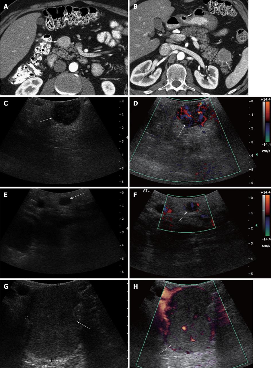
Figure 8 A 46-year-old male with multiple endocrine neoplasia type I syndrome and pancreatic neuroendocrine tumor.
A, B: Axial contrast-enhanced computed tomography (CT) images show hypervascular masses in the head and tail of the pancreas, consistent with neuroendocrine tumors; C: Intraoperative ultrasound image shows a well-defined solid mass in the head of the pancreas, consistent with the neuroendocrine tumor on CT; D: Doppler interrogation reveals increased vascularity within this mass; E, F: Intraoperative grayscale and color Doppler ultrasound images detect a 5 mm solid mass with internal vascularity in the head of the pancreas, consistent with a small neuroendocrine tumor. This lesion was not identified on the CT examination; G, H: Intraoperative grayscale and color Doppler ultrasound reveals the large dominant mass in the tail of the pancreas, consistent with a neuroendocrine tumor.
- Citation: Marcal LP, Patnana M, Bhosale P, Bedi DG. Intraoperative abdominal ultrasound in oncologic imaging. World J Radiol 2013; 5(3): 51-60
- URL: https://www.wjgnet.com/1949-8470/full/v5/i3/51.htm
- DOI: https://dx.doi.org/10.4329/wjr.v5.i3.51


