Copyright
©2013 Baishideng.
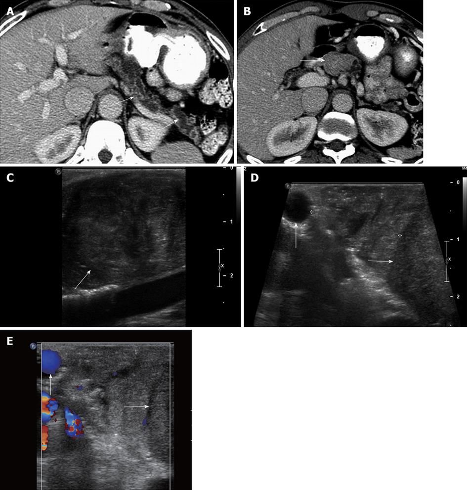
Figure 7 A 60-year-old male with pancreatic duct dilatation and suspected mass pancreatic head mass.
A: Axial contrast-enhanced computed tomography (CT) images show marked pancreatic ductal dilatation in the tail and body of the pancreas, with atrophy of the pancreatic parenchyma; B: Axial contrast-enhanced CT shows fullness in the region of the head of the pancreas, concerning for an isodense pancreatic head mass; C: Intraoperative ultrasound image clearly defines a solid mass in the head of the pancreas, consistent with a pancreatic ductal adenocarcinoma; D, E: Grayscale (D) and color Doppler (E) intraoperative ultrasound images show to better advantage the margins of the mass (arrow) in the cephalad head of the pancreas. The lesion is confined to the pancreas, and does not involve the gastroduodenal artery (vertical arrow). Intraoperative ultrasound is useful to assess size, margins of pancreatic mass, and their relationship with the adjacent vessels. This is particularly useful in cases of isodense pancreatic masses, which can be very difficult to evaluate with CT.
- Citation: Marcal LP, Patnana M, Bhosale P, Bedi DG. Intraoperative abdominal ultrasound in oncologic imaging. World J Radiol 2013; 5(3): 51-60
- URL: https://www.wjgnet.com/1949-8470/full/v5/i3/51.htm
- DOI: https://dx.doi.org/10.4329/wjr.v5.i3.51


