Copyright
©2013 Baishideng.
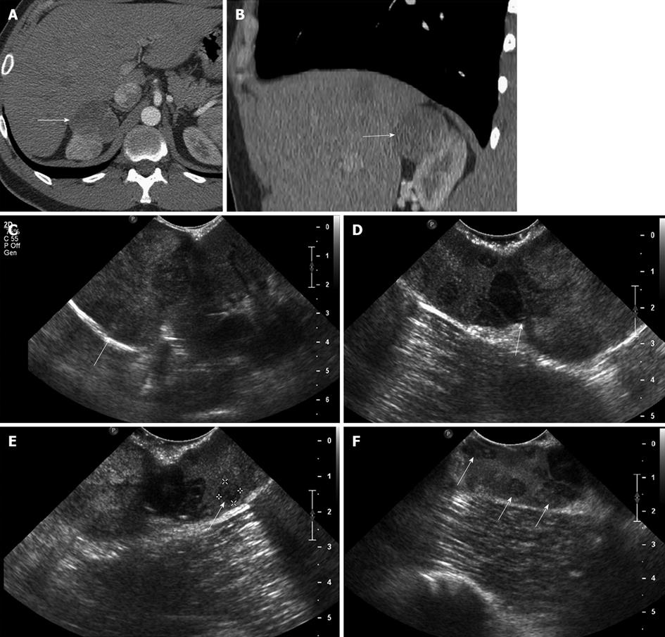
Figure 6 A 29-year-old male with renal cell carcinoma of the right kidney.
A, B: Axial (A) and coronal (B) contrast-enhanced computed tomography image show a large solid mass in the superior pole of the right kidney, consistent with a renal cell carcinoma; C: Intraoperative longitudinal sonogram shows an exophytic large solid mass in the superior pole of the right kidney; D: Intraoperative ultrasound image shows the point of attachment of the solid mass to the superior pole of the right kidney; E, F: High resolution intraoperative ultrasound image detects additional subcentimeter solid renal masses in the superior pole of the right kidney, near the site of origin of the large exophytic right renal mass. These findings alerted the surgeon that a deeper resection needed to be performed in order to secure tumor-free resection margin.
- Citation: Marcal LP, Patnana M, Bhosale P, Bedi DG. Intraoperative abdominal ultrasound in oncologic imaging. World J Radiol 2013; 5(3): 51-60
- URL: https://www.wjgnet.com/1949-8470/full/v5/i3/51.htm
- DOI: https://dx.doi.org/10.4329/wjr.v5.i3.51


