Copyright
©2013 Baishideng.
World J Radiol. Mar 28, 2013; 5(3): 126-142
Published online Mar 28, 2013. doi: 10.4329/wjr.v5.i3.126
Published online Mar 28, 2013. doi: 10.4329/wjr.v5.i3.126
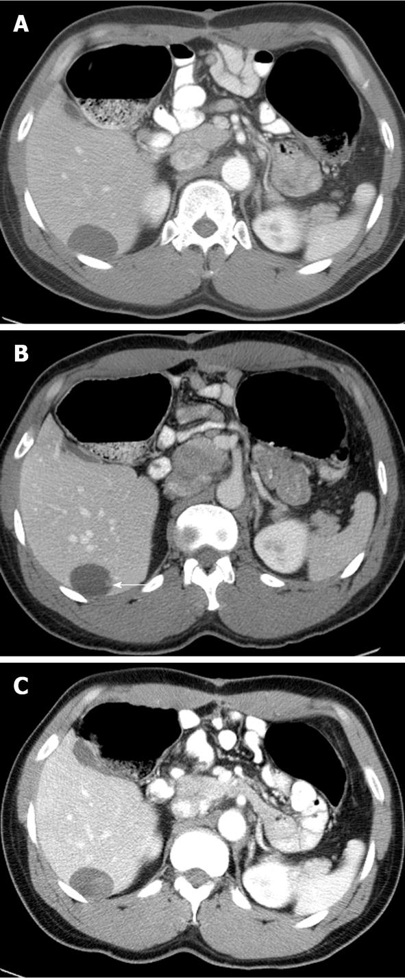
Figure 7 Axial computed tomography images of the same patient in Figure 6 at a later time showed progression of metastatic disease.
Immediately following the second course of targeted therapy, the hepatic metastasis showed homogenous hypoenhancement compatible with treatment response (A). A small nodular enhancing focus within the lesion is the radiographic finding of recurrent disease (arrow in B) which had progressed and appeared more conspicuous (C).
- Citation: Peungjesada S, Chuang HH, Prasad SR, Choi H, Loyer EM, Bronstein Y. Evaluation of cancer treatment in the abdomen: Trends and advances. World J Radiol 2013; 5(3): 126-142
- URL: https://www.wjgnet.com/1949-8470/full/v5/i3/126.htm
- DOI: https://dx.doi.org/10.4329/wjr.v5.i3.126


