Copyright
©2013 Baishideng.
World J Radiol. Mar 28, 2013; 5(3): 126-142
Published online Mar 28, 2013. doi: 10.4329/wjr.v5.i3.126
Published online Mar 28, 2013. doi: 10.4329/wjr.v5.i3.126
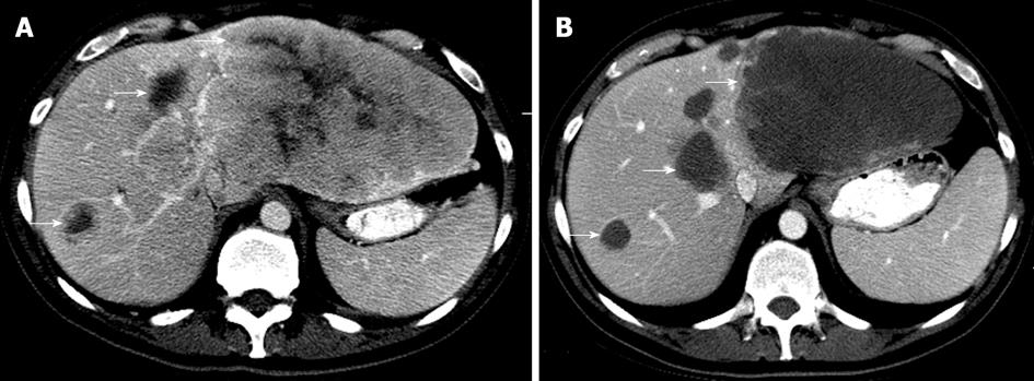
Figure 2 Axial computed tomography images in the portovenous phase of a 50-year-old female with colorectal liver metastases.
A: Pretreatment evaluation showed multiple heterogeneously enhancing hepatic masses that are compatible with known hepatic metastases. The entire left hepatic lobe was occupied by the masses. Note the thickened nodular interface between the mass and normal liver (arrow); B: Liver metastases became more homogenous and showed well-defined interfaces with the normal liver parenchyma after Folfox and Avastin, corresponding to treatment response. Note the well defined interface (arrows).
- Citation: Peungjesada S, Chuang HH, Prasad SR, Choi H, Loyer EM, Bronstein Y. Evaluation of cancer treatment in the abdomen: Trends and advances. World J Radiol 2013; 5(3): 126-142
- URL: https://www.wjgnet.com/1949-8470/full/v5/i3/126.htm
- DOI: https://dx.doi.org/10.4329/wjr.v5.i3.126


