Copyright
©2013 Baishideng.
World J Radiol. Mar 28, 2013; 5(3): 113-125
Published online Mar 28, 2013. doi: 10.4329/wjr.v5.i3.113
Published online Mar 28, 2013. doi: 10.4329/wjr.v5.i3.113
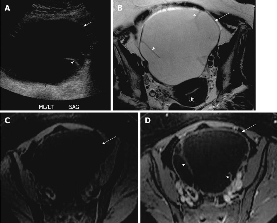
Figure 8 Mucinous cystadenoma.
A 44-year-old woman with large pelvic mass. A: Transabdominal pelvic ultrasound demonstrates a large cystic lesion (arrow) in the mid pelvis with a thin septation (arrowhead); B-D: Axial T2 weighted image (B), axial pre contrast (C) and fat saturated post contrast T1 weighted image (D), demonstrates a large left ovarian cystic lesion (arrow) with several thin septations (arrowheads). The lesion has a low signal intensity on T1 weighted image and high signal on T2 weighted image and hypointense septae, with no significant post contrast enhancement. Ut: Uterus.
- Citation: Wasnik AP, Menias CO, Platt JF, Lalchandani UR, Bedi DG, Elsayes KM. Multimodality imaging of ovarian cystic lesions: Review with an imaging based algorithmic approach. World J Radiol 2013; 5(3): 113-125
- URL: https://www.wjgnet.com/1949-8470/full/v5/i3/113.htm
- DOI: https://dx.doi.org/10.4329/wjr.v5.i3.113


