Copyright
©2013 Baishideng.
World J Radiol. Mar 28, 2013; 5(3): 113-125
Published online Mar 28, 2013. doi: 10.4329/wjr.v5.i3.113
Published online Mar 28, 2013. doi: 10.4329/wjr.v5.i3.113
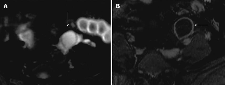
Figure 3 Functional cyst on magnetic resonance imaging.
A 31-year-old woman with magnetic resonance abdomen pelvis performed for inflammatory bowel disease evaluation. A: Axial fat suppressed T2 weighted image with fat saturation demonstrates high signal intensity focus in the left ovary (arrow), without any internal debris or septation; B: Axial post contrast fat suppressed T1 weighted image showing thin enhancing wall of the cyst without internal enhancement or septation (arrow).
- Citation: Wasnik AP, Menias CO, Platt JF, Lalchandani UR, Bedi DG, Elsayes KM. Multimodality imaging of ovarian cystic lesions: Review with an imaging based algorithmic approach. World J Radiol 2013; 5(3): 113-125
- URL: https://www.wjgnet.com/1949-8470/full/v5/i3/113.htm
- DOI: https://dx.doi.org/10.4329/wjr.v5.i3.113


