Copyright
©2013 Baishideng Publishing Group Co.
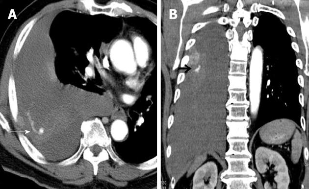
Figure 1 Contrast-enhanced computed tomography shows massive right hemothorax, tumor stain, and extravasation of contrast agent (arrow) in the right chest wall.
- Citation: Nagao E, Hirakawa M, Soeda H, Tsuruta S, Sakai H, Honda H. Transcatheter arterial embolization for chest wall metastasis of hepatocellular carcinoma. World J Radiol 2013; 5(2): 45-48
- URL: https://www.wjgnet.com/1949-8470/full/v5/i2/45.htm
- DOI: https://dx.doi.org/10.4329/wjr.v5.i2.45


