Copyright
©2013 Baishideng Publishing Group Co.
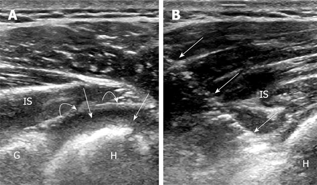
Figure 2 Ultrasound image through the posterior shoulder.
A: Axial ultrasound image through the posterior shoulder shows a large joint effusion (straight arrows) with fluid between the humeral head (H) and the adjacent posterior capsule (curved arrows); B: Ultrasound guided aspiration of the posterior shoulder. Note the needle (arrows). Cultures grew Streptococcus crista. IS: infraspinatus; G: Posterior glenoid.
- Citation: Vollman AT, Craig JG, Hulen R, Ahmed A, Zervos MJ, Holsbeeck MV. Review of three magnetic resonance arthrography related infections. World J Radiol 2013; 5(2): 41-44
- URL: https://www.wjgnet.com/1949-8470/full/v5/i2/41.htm
- DOI: https://dx.doi.org/10.4329/wjr.v5.i2.41


