Copyright
©2013 Baishideng Publishing Group Co.
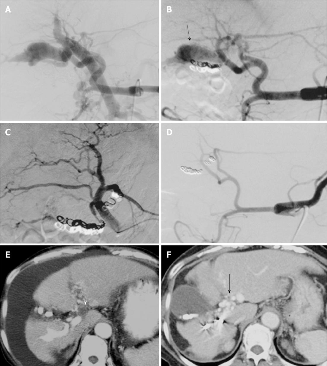
Figure 1 Case 2: A 60-year-old woman with arterioportal fistula and refractory ascites related to liver biopsy performed 5 years earlier.
A: Celiac arteriogram before transcatheter arterial embolization (TAE) shows enlarged, tortuous hepatic artery and immediate retrograde visualization of main portal vein (PV) trunk. Note branches of the right hepatic artery (A7 and A8) supplying the fistulous connection to the right PV branch. A7 was superselectively embolized using microcoils; B: Celiac arteriogram 10 d after the initial TAE shows a peripheral branch of the right PV (arrow). Additional TAE for A6 was performed using microcoils; C: Right hepatic arteriogram after second TAE indicates obliteration of arterioportal fistula (APF); D: Follow-up celiac arteriogram 12 mo after TAE shows obliteration of APF and improved enlarged hepatic artery; E: Qualifying computed tomography (CT) (hepatic arterial phase image) before TAE. CT shows simultaneous enhancement of the aorta and right branch of PV (white arrow), indicating severe APF, PV thrombi in the umbilical portion (arrowhead) and moderate ascites; F: Follow-up CT (portal venous phase image) 50 mo after TAE. CT shows the obliteration of APF, the disappearance of ascites and cavernous transformation (arrow).
- Citation: Hirakawa M, Nishie A, Asayama Y, Ishigami K, Ushijima Y, Fujita N, Honda H. Clinical outcomes of symptomatic arterioportal fistulas after transcatheter arterial embolization. World J Radiol 2013; 5(2): 33-40
- URL: https://www.wjgnet.com/1949-8470/full/v5/i2/33.htm
- DOI: https://dx.doi.org/10.4329/wjr.v5.i2.33


