Copyright
©2013 Baishideng Publishing Group Co.
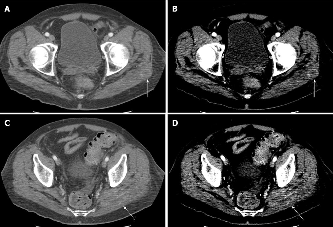
Figure 7 Skeletal muscle metastasis.
Axial sections from contrast enhanced computed tomography (CT) examinations in two different patients with metastases to the gluteus maximus (arrows). A, C: Images are displayed in typical soft tissue windows (ww/wl: 500/50); B, D: Images are the same acquired images displayed in more narrow windows (ww/wl: 200/50). Note the increased conspicuity of the lesions when narrow windows are used.
- Citation: Trout AT, Rabinowitz RS, Platt JF, Elsayes KM. Melanoma metastases in the abdomen and pelvis: Frequency and patterns of spread. World J Radiol 2013; 5(2): 25-32
- URL: https://www.wjgnet.com/1949-8470/full/v5/i2/25.htm
- DOI: https://dx.doi.org/10.4329/wjr.v5.i2.25


