Copyright
©2013 Baishideng Publishing Group Co.
World J Radiol. Dec 28, 2013; 5(12): 498-502
Published online Dec 28, 2013. doi: 10.4329/wjr.v5.i12.498
Published online Dec 28, 2013. doi: 10.4329/wjr.v5.i12.498
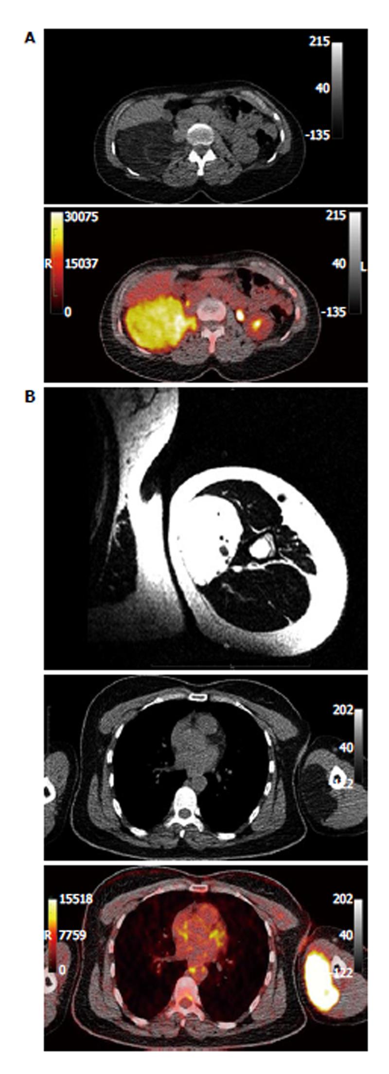
Figure 1 Computed tomography and magnetic resonance imaging.
A: Computed tomography (upper row) and fused positron emission tomography/CT (PET/CT) (lower row) images of the retroperitoneal tumor; B: Magnetic resonance imaging (upper row), CT (middle row) and fused positron emission tomography/CT (PET/CT) (lower low) images of the tumor located in the left upper arm. Magnetic resonance imaging demonstrates a well-circumscribed mass without infiltration of the surrounding tissues. CT reveals a fat-equivalent mass, while PET/CT demonstrates intense fluorodeoxyglucose accumulation in the tumor area. CT: Computed tomography.
- Citation: Sachpekidis C, Roumia S, Schwarzbach M, Dimitrakopoulou-Strauss A. Dynamic 18F-fluorodeoxyglucose positron emission tomography/CT in hibernoma: Enhanced tracer uptake mimicking liposarcoma. World J Radiol 2013; 5(12): 498-502
- URL: https://www.wjgnet.com/1949-8470/full/v5/i12/498.htm
- DOI: https://dx.doi.org/10.4329/wjr.v5.i12.498


