Copyright
©2013 Baishideng Publishing Group Co.
World J Radiol. Dec 28, 2013; 5(12): 472-483
Published online Dec 28, 2013. doi: 10.4329/wjr.v5.i12.472
Published online Dec 28, 2013. doi: 10.4329/wjr.v5.i12.472
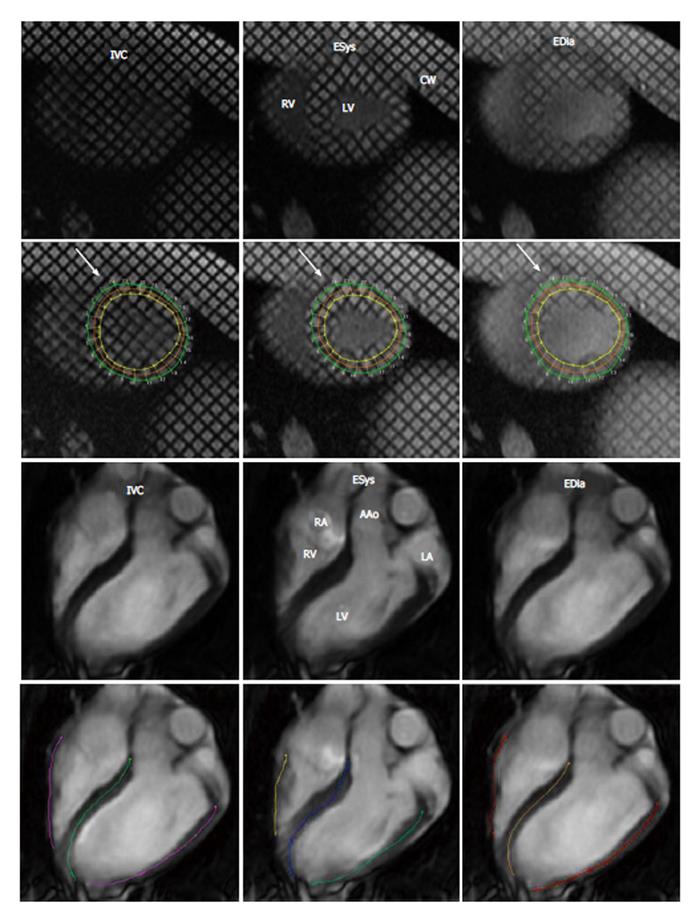
Figure 1 Representative tagged and cine magnetic resonance images with tracing method.
Top row demonstrates short-axis and long-axis magnetic resonance images (MRI) images, while bottom row demonstrates images after tracing of the myocardium using HARP. Left three columns are cine tagged MRI and right three columns are cine MRI. IVC: Isovolumetric contraction; ESys: End systole; EDia: End diastole; LV: Left ventricle; RV: Right ventricle; CW: Chest wall; RA: Right atrium; LA: Left atrium; AAo: Ascending aorta.
- Citation: Suhail MS, Wilson MW, Hetts SW, Saeed M. Magnetic resonance imaging characterization of circumferential and longitudinal strain under various coronary interventions in swine. World J Radiol 2013; 5(12): 472-483
- URL: https://www.wjgnet.com/1949-8470/full/v5/i12/472.htm
- DOI: https://dx.doi.org/10.4329/wjr.v5.i12.472


