Copyright
©2013 Baishideng Publishing Group Co.
World J Radiol. Dec 28, 2013; 5(12): 460-467
Published online Dec 28, 2013. doi: 10.4329/wjr.v5.i12.460
Published online Dec 28, 2013. doi: 10.4329/wjr.v5.i12.460
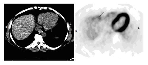
Figure 4 Noise artifact in the liver.
A 42-year-old man with history of cervical cancer had fluorodeoxyglucose positron emission tomography-computer tomography (PET-CT) for initial staging. Axial PET image of the upper abdomen shows irregular uptake in the anterior margin (arrow) but without discrete lesions on an integrated CT, representing noise artifacts. Subsequent diagnostic CT was negative.
- Citation: Liu Y. Fluorodeoxyglucose uptake in absence of CT abnormality on PET-CT: What is it? World J Radiol 2013; 5(12): 460-467
- URL: https://www.wjgnet.com/1949-8470/full/v5/i12/460.htm
- DOI: https://dx.doi.org/10.4329/wjr.v5.i12.460


