Copyright
©2013 Baishideng Publishing Group Co.
World J Radiol. Nov 28, 2013; 5(11): 421-429
Published online Nov 28, 2013. doi: 10.4329/wjr.v5.i11.421
Published online Nov 28, 2013. doi: 10.4329/wjr.v5.i11.421
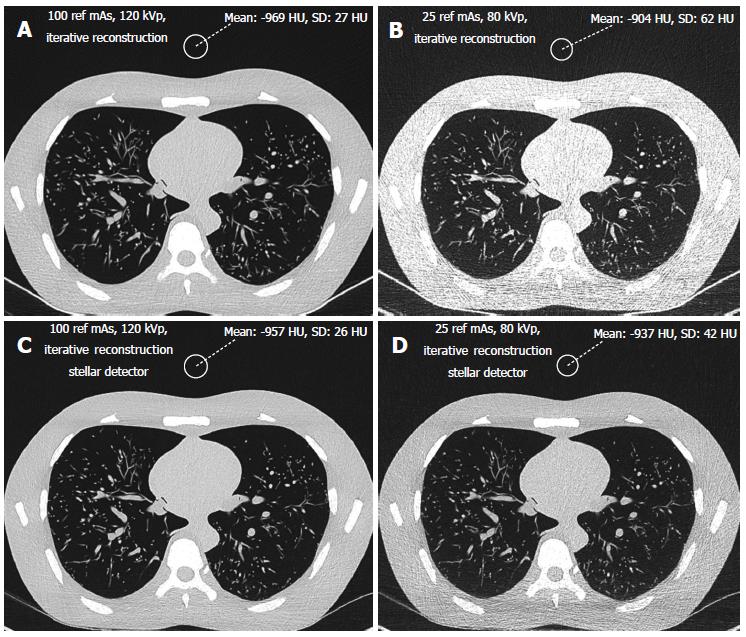
Figure 1 Imaging of the lung using iterative reconstruction with (C, D) and without (A, B) the Stellar detector.
The chest phantom was scanned at a standard dose level with a 100 reference mAs tube current time and a 120 kVp voltage (A, C) and the lowest dose level of 25 ref mAs and 80 kVp. At the standard dose level both images with and without the Stellar detector (A,C) had similar noise levels (20-30 HU), while at the lowest dose, the image quality was obviously better with the Stellar detector (D).
- Citation: Christe A, Heverhagen J, Ozdoba C, Weisstanner C, Ulzheimer S, Ebner L. CT dose and image quality in the last three scanner generations. World J Radiol 2013; 5(11): 421-429
- URL: https://www.wjgnet.com/1949-8470/full/v5/i11/421.htm
- DOI: https://dx.doi.org/10.4329/wjr.v5.i11.421


