Copyright
©2013 Baishideng Publishing Group Co.
World J Radiol. Nov 28, 2013; 5(11): 411-420
Published online Nov 28, 2013. doi: 10.4329/wjr.v5.i11.411
Published online Nov 28, 2013. doi: 10.4329/wjr.v5.i11.411
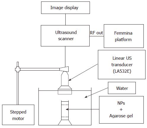
Figure 2 Scheme of the employed ultrasound acquisition system.
The image display and the linear ultrasound transducer (indicated by the arrow in the figure) are electronically connected to the ultrasound scanner; the radiofrequency signal output is transmitted to the FEMMINA Platform by means of optic fiber. The set up is completed by a nanoparticle (NP) containing phantom (indicated in figure as “NP + agarose gel”) immersed in a water tank (pointed by the respective arrow). US: Ultrasound.
- Citation: Chiriacò F, Soloperto G, Greco A, Conversano F, Ragusa A, Menichetti L, Casciaro S. Magnetically-coated silica nanospheres for dual-mode imaging at low ultrasound frequency. World J Radiol 2013; 5(11): 411-420
- URL: https://www.wjgnet.com/1949-8470/full/v5/i11/411.htm
- DOI: https://dx.doi.org/10.4329/wjr.v5.i11.411


