Copyright
©2013 Baishideng Publishing Group Co.
World J Radiol. Nov 28, 2013; 5(11): 386-397
Published online Nov 28, 2013. doi: 10.4329/wjr.v5.i11.386
Published online Nov 28, 2013. doi: 10.4329/wjr.v5.i11.386
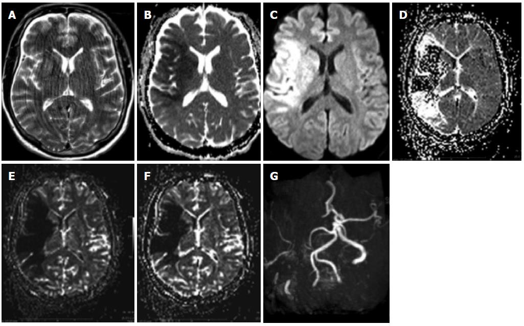
Figure 6 Patient with penumbra and mismatch.
A: A patient with an acute left-sided hemisyndrome was referred to our hospital: T2 images showed no clear signs of ischemia despite motion artifacts; B, C: The apparent diffusion coefficient (ADC) was lowered in the right MCA territory with a corresponding hyperintensity on the diffusion-weighted imaging (DWI) images with the maximum b value; D-F: There was additional hypoperfusion in a more extensive area on the MTT maps (D) as well as on the maps of rCBV (E) and RCB (F), which was sue to an occlusion of the MCA on the right.
- Citation: Pereira VM, Vargas MI, Marcos A, Bijlenga P, Narata AP, Haller S, Lövblad KO. Diagnostic neuroradiology for the interventional neuroradiologist. World J Radiol 2013; 5(11): 386-397
- URL: https://www.wjgnet.com/1949-8470/full/v5/i11/386.htm
- DOI: https://dx.doi.org/10.4329/wjr.v5.i11.386


