Copyright
©2013 Baishideng Publishing Group Co.
World J Radiol. Nov 28, 2013; 5(11): 386-397
Published online Nov 28, 2013. doi: 10.4329/wjr.v5.i11.386
Published online Nov 28, 2013. doi: 10.4329/wjr.v5.i11.386
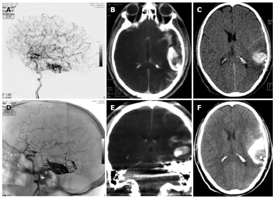
Figure 4 Dual energy computed tomography.
A, D: Patient having undergone angiography and embolization for a temporal left AVM; B, E: On the immediate expert computed tomography (CT) performed on the angio table, there was a large area of hyperdensity with probably contrast and blood but which were not distinguishable from one another: the Dual source CT shows that this is mainly due to contrast since there is sonly a small area of blood surrounding the embolized material; C, F: Again a normal C CT afterwards shows the more extensive hyperdensity in the subarachnoid regions due to contrast and blood.
- Citation: Pereira VM, Vargas MI, Marcos A, Bijlenga P, Narata AP, Haller S, Lövblad KO. Diagnostic neuroradiology for the interventional neuroradiologist. World J Radiol 2013; 5(11): 386-397
- URL: https://www.wjgnet.com/1949-8470/full/v5/i11/386.htm
- DOI: https://dx.doi.org/10.4329/wjr.v5.i11.386


