Copyright
©2013 Baishideng Publishing Group Co.
World J Radiol. Nov 28, 2013; 5(11): 386-397
Published online Nov 28, 2013. doi: 10.4329/wjr.v5.i11.386
Published online Nov 28, 2013. doi: 10.4329/wjr.v5.i11.386
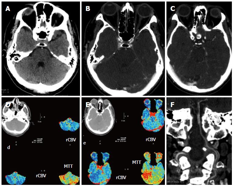
Figure 3 Patient with symptoms referable to the posterior fossa.
A-C: Unenhanced computed tomography (CT) shows a possible hyperdense artery sign of the basilar artery (A) which is confirmed by CT angiography: the basilar artery enhances less than the rest of the vessels (B, C); D, E: Perfusion imaging shows a drop in hemodynamics in the posterior fossa; F: The coronal reconstruction of the angio-CT shows well the length of the thrombus.
- Citation: Pereira VM, Vargas MI, Marcos A, Bijlenga P, Narata AP, Haller S, Lövblad KO. Diagnostic neuroradiology for the interventional neuroradiologist. World J Radiol 2013; 5(11): 386-397
- URL: https://www.wjgnet.com/1949-8470/full/v5/i11/386.htm
- DOI: https://dx.doi.org/10.4329/wjr.v5.i11.386


