Copyright
©2013 Baishideng Publishing Group Co.
World J Radiol. Oct 28, 2013; 5(10): 372-380
Published online Oct 28, 2013. doi: 10.4329/wjr.v5.i10.372
Published online Oct 28, 2013. doi: 10.4329/wjr.v5.i10.372
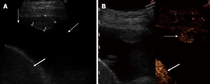
Figure 3 Delayed arterial enhancement.
A: Tranverse sonogram of the left chest wall in the seventh intercostal space along the mid-axillary line showing an isoechoic pleural nodule, pleural effusion (thin arrows), and spleen (large arrow); B: Transverse contrast-enhanced sonogram in the arterial phase showing inhomogeneous enhancement of the nodule (thin arrow) that occurs contemporaneously to the enhancement of the spleen (large arrow) (left side of the split-screen); the enhancement of the nodule is less marked than that of the spleen. Final diagnosis was pleural metastasis from colon cancer.
- Citation: Sartori S, Postorivo S, Vece FD, Ermili F, Tassinari D, Tombesi P. Contrast-enhanced ultrasonography in peripheral lung consolidations: What’s its actual role? World J Radiol 2013; 5(10): 372-380
- URL: https://www.wjgnet.com/1949-8470/full/v5/i10/372.htm
- DOI: https://dx.doi.org/10.4329/wjr.v5.i10.372


