Copyright
©2013 Baishideng Publishing Group Co.
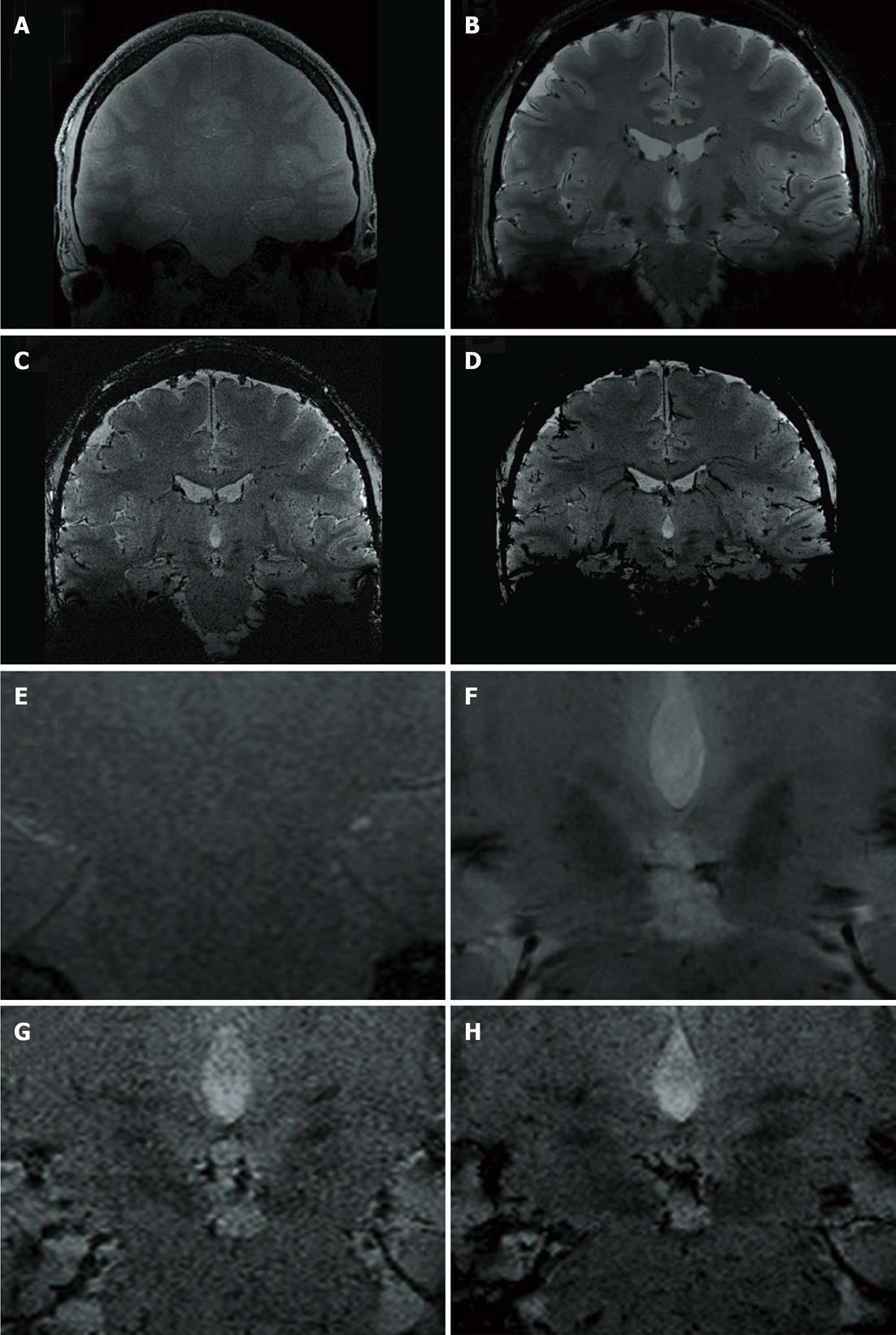
Figure 2 Non-enlarged (A-D) and enlarged (E-H) representative coronal images at the level of the zona incerta.
A, E: T1-weighted gradient-echo coronal; B, F: FLASH2D-T2Star coronal; C, G: Susceptibility-weighted imaging (SWI) coronal; D, H: SWI-minimum intensity projections coronal.
- Citation: Kerl HU, Gerigk L, Brockmann MA, Huck S, Al-Zghloul M, Groden C, Hauser T, Nagel AM, Nölte IS. Imaging for deep brain stimulation: The zona incerta at 7 Tesla. World J Radiol 2013; 5(1): 5-16
- URL: https://www.wjgnet.com/1949-8470/full/v5/i1/5.htm
- DOI: https://dx.doi.org/10.4329/wjr.v5.i1.5


