Copyright
©2013 Baishideng Publishing Group Co.
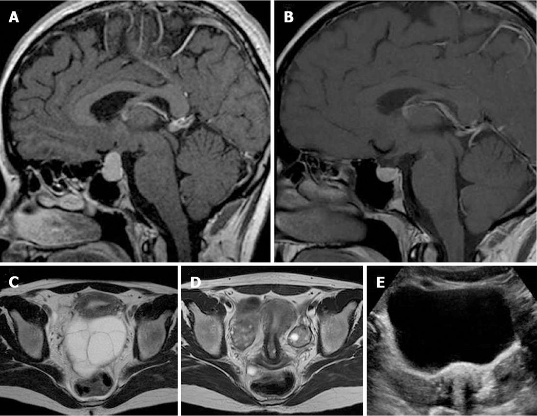
Figure 4 Sellar sagittal MR images with gadolinium administration before (A) and after (B) thyroid hormone replacement therapy and pelvic axial T2-weighted images before (C) and after (D) thyroid hormone replacement therapy revealed regression of the pituitary mass and ovarian cystic enlargement of both ovaries.
E: A corresponding transabdominal ultrasound image after thyroid therapy demonstrating regression of both ovarian cystic masses.
- Citation: Kanza RE, Gagnon S, Villeneuve H, Laverdiere D, Rousseau I, Bordeleau E, Berube M. Spontaneous ovarian hyperstimulation syndrome and pituitary hyperplasia mimicking macroadenoma associated with primary hypothyroidism. World J Radiol 2013; 5(1): 20-24
- URL: https://www.wjgnet.com/1949-8470/full/v5/i1/20.htm
- DOI: https://dx.doi.org/10.4329/wjr.v5.i1.20


