Copyright
©2012 Baishideng Publishing Group Co.
World J Radiol. Sep 28, 2012; 4(9): 391-400
Published online Sep 28, 2012. doi: 10.4329/wjr.v4.i9.391
Published online Sep 28, 2012. doi: 10.4329/wjr.v4.i9.391
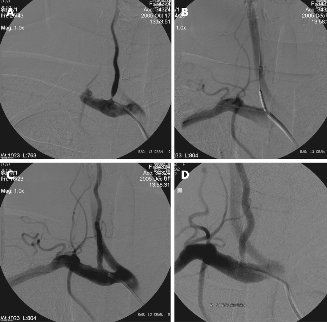
Figure 4 Radiological follow-up.
A: Right subclavian angiography posterior-anterior (PA) view shows significant stenosis of the right vertebral artery origin; B: Placement of the coronary balloon-expandable stent in the stenosed segment; C: After opening of the stent angiography shows well opposition of the stent and lack of residual stenosis; D: Eighteen months control angiography shows patency of the right VA origin and stent.
- Citation: Kocak B, Korkmazer B, Islak C, Kocer N, Kizilkilic O. Endovascular treatment of extracranial vertebral artery stenosis. World J Radiol 2012; 4(9): 391-400
- URL: https://www.wjgnet.com/1949-8470/full/v4/i9/391.htm
- DOI: https://dx.doi.org/10.4329/wjr.v4.i9.391


