Copyright
©2012 Baishideng Publishing Group Co.
World J Radiol. Aug 28, 2012; 4(8): 379-386
Published online Aug 28, 2012. doi: 10.4329/wjr.v4.i8.379
Published online Aug 28, 2012. doi: 10.4329/wjr.v4.i8.379
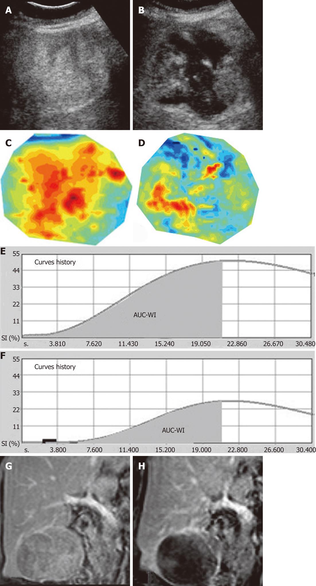
Figure 4 Another case of hepatocellular carcinoma studied with non-parametric contrast-enhanced ultrasonography and parametric contrast-enhanced ultrasound before and after the first session of transarterial chemoembolization.
A, B: Non-parametric contrast-enhanced ultrasonography (npCEUS) at the plane of longest tumor diameter prior to (A) and post transarterial chemoembolization (TACE) (B), shows non-enhancing (necrotic) areas post-TACE as well as multiple residual enhancing components; C, D: parametric images prior to (C) and post TACE (D) depict well-perfused areas with red and yellow tones, while hypoperfused areas are depicted with tones of blue; E, F: Time-intensity curves prior to (E) and post TACE (F), confirm a reduction in peak intensity (PI) and area under the curve during wash-in (AUC-WI) after TACE; G, H: Corresponding coronal, T1-weighted, gadolinium-enhanced magnetic resonance images of the same tumor, prior to (G) and post TACE (H) correlate favorably with contrast-enhanced ultrasonography images and confirm the significant reduction in lesional enhancement, but no tumor shrinkage. SI: Signal intensity.
- Citation: Moschouris H, Malagari K, Marinis A, Kornezos I, Stamatiou K, Nikas G, Papadaki MG, Gkoutzios P. Hepatocellular carcinoma treated with transarterial chemoembolization: Evaluation with parametric contrast-enhanced ultrasonography. World J Radiol 2012; 4(8): 379-386
- URL: https://www.wjgnet.com/1949-8470/full/v4/i8/379.htm
- DOI: https://dx.doi.org/10.4329/wjr.v4.i8.379


