Copyright
©2012 Baishideng Publishing Group Co.
World J Radiol. Aug 28, 2012; 4(8): 353-371
Published online Aug 28, 2012. doi: 10.4329/wjr.v4.i8.353
Published online Aug 28, 2012. doi: 10.4329/wjr.v4.i8.353
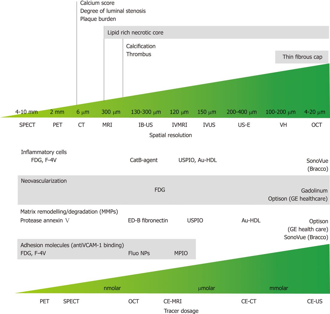
Figure 10 Spatial resolution and dose-effectiveness of different diagnostic imaging modalities.
Minimum spatial resolution required for the identification of the indicated morphological feature of vulnerable atherosclerotic plaque (top); dose effectiveness in the identification of specific biological processes and compound; specific target and respective tracer are indicated (bottom). SPECT: Single photon emission computed tomography; PET: Positron emission tomography; CT: Computed tomography; MRI: Magnetic resonance imaging; IB-US: Integrated backscatter ultrasonography; IVMRI: Intravascular magnetic resonance imaging; IVUS: Intravascular ultrasound; US-E: Intravascular ultrasound employed in elastography; VH: Virtual histology; OCT: Optical coherence tomography; CE-MRI: Contrast-enhanced magnetic resonance imaging; CE-CT: Contrast-enhanced computed tomography; CE-US: Contrast-enhanced ultrasonography; FDG: Fluorodeoxyglucose; USPIO: Ultra-small super paramagnetic iron oxide; Au-HDL: High-density lipoprotein nanoparticle contrast agent; VCAM: Vascular cell adhesion molecule; NP: Nanoparticle; MPIO: Microparticles of iron oxide.
- Citation: Soloperto G, Casciaro S. Progress in atherosclerotic plaque imaging. World J Radiol 2012; 4(8): 353-371
- URL: https://www.wjgnet.com/1949-8470/full/v4/i8/353.htm
- DOI: https://dx.doi.org/10.4329/wjr.v4.i8.353


