Copyright
©2012 Baishideng Publishing Group Co.
World J Radiol. Aug 28, 2012; 4(8): 353-371
Published online Aug 28, 2012. doi: 10.4329/wjr.v4.i8.353
Published online Aug 28, 2012. doi: 10.4329/wjr.v4.i8.353
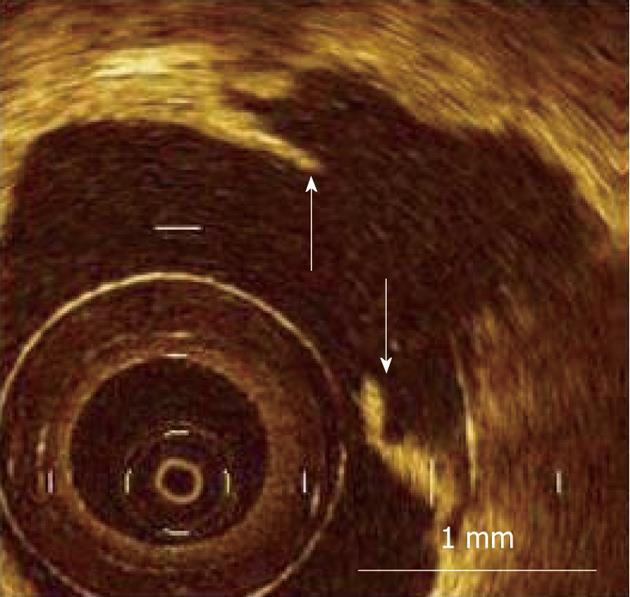
Figure 7 Measurement of fibrous cap thickness in ruptured plaque using optical coherence tomography.
Residual fibrous cap was identified as a flap between the lumen of the coronary artery and the cavity of plaque, and its thickness was measured at the thinnest part (arrows). Scale bar = 1 mm (courtesy of Dr. Kubo).
- Citation: Soloperto G, Casciaro S. Progress in atherosclerotic plaque imaging. World J Radiol 2012; 4(8): 353-371
- URL: https://www.wjgnet.com/1949-8470/full/v4/i8/353.htm
- DOI: https://dx.doi.org/10.4329/wjr.v4.i8.353


