Copyright
©2012 Baishideng Publishing Group Co.
World J Radiol. Aug 28, 2012; 4(8): 353-371
Published online Aug 28, 2012. doi: 10.4329/wjr.v4.i8.353
Published online Aug 28, 2012. doi: 10.4329/wjr.v4.i8.353
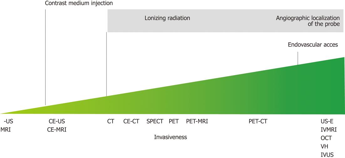
Figure 2 Illustration of the invasiveness of the possible plaque imaging modalities along with the reasons of their grading.
MRI: Magnetic resonance imaging; IB-US: Integrated backscatter ultrasound; CE-US: Contrast enhanced ultrasound; CE-MRI: Contrast enhanced magnetic resonance imaging; CT: Computed tomography; CE-CT: Contrast enhanced computed tomography; SPECT: Single proton emission computed tomography; PET: Positron emission tomography; US-E: Ultrasound elastography; IVMRI: Intravascular magnetic resonance imaging; OCT: Optical coherence tomography; VH: Virtual histology; IVUS: Intravascular ultrasound.
- Citation: Soloperto G, Casciaro S. Progress in atherosclerotic plaque imaging. World J Radiol 2012; 4(8): 353-371
- URL: https://www.wjgnet.com/1949-8470/full/v4/i8/353.htm
- DOI: https://dx.doi.org/10.4329/wjr.v4.i8.353


