Copyright
©2012 Baishideng Publishing Group Co.
World J Radiol. Aug 28, 2012; 4(8): 353-371
Published online Aug 28, 2012. doi: 10.4329/wjr.v4.i8.353
Published online Aug 28, 2012. doi: 10.4329/wjr.v4.i8.353
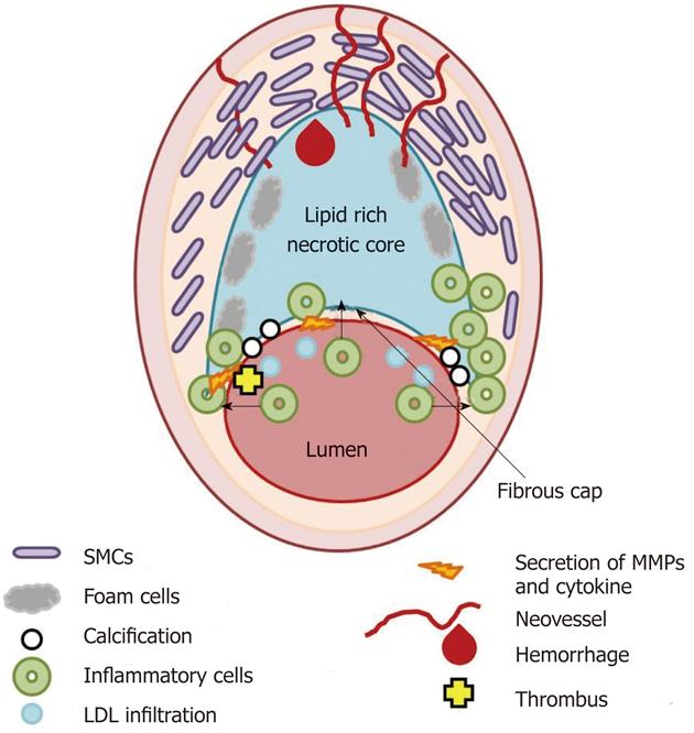
Figure 1 Scheme of the thin fibrous cap atheroma.
Main cellular components characterizing atherosclerotic plaque formation and destabilization are illustrated as well as biological and morphological features occurring in vulnerable plaque. SMCs: Smooth muscle cells; LDL: Low density lipoprotein; MMPs: Matrix metalloproteases.
- Citation: Soloperto G, Casciaro S. Progress in atherosclerotic plaque imaging. World J Radiol 2012; 4(8): 353-371
- URL: https://www.wjgnet.com/1949-8470/full/v4/i8/353.htm
- DOI: https://dx.doi.org/10.4329/wjr.v4.i8.353


