Copyright
©2012 Baishideng Publishing Group Co.
World J Radiol. Aug 28, 2012; 4(8): 345-352
Published online Aug 28, 2012. doi: 10.4329/wjr.v4.i8.345
Published online Aug 28, 2012. doi: 10.4329/wjr.v4.i8.345
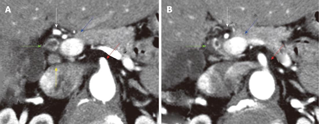
Figure 9 Distal bile duct tumors.
Post-contrast computed tomography (CT) examination of the abdomen in a 58-year-old female with cholangiocarcinoma of the bile duct; A: CT image at a cranial location shows dilatation of the bile duct and bile duct enhancement (green arrow); B: CT image at a caudal location shows enhancement of the distal bile duct. There is a small regional lymph node (yellow arrow). The hepatic artery (white arrow), portal vein (blue arrow), and superior mesenteric artery (red arrow) are spared. Radiologically, appearances are consistent with a T1 tumor (periductal fat not involved) and this was confirmed by pathology.
- Citation: Ganeshan D, Moron FE, Szklaruk J. Extrahepatic biliary cancer: New staging classification. World J Radiol 2012; 4(8): 345-352
- URL: https://www.wjgnet.com/1949-8470/full/v4/i8/345.htm
- DOI: https://dx.doi.org/10.4329/wjr.v4.i8.345


