Copyright
©2012 Baishideng Publishing Group Co.
World J Radiol. Aug 28, 2012; 4(8): 345-352
Published online Aug 28, 2012. doi: 10.4329/wjr.v4.i8.345
Published online Aug 28, 2012. doi: 10.4329/wjr.v4.i8.345
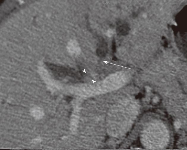
Figure 7 T4 - proximal bile tumor.
Post-contrast computed tomography exam at the level of the intrahepatic bile duct in a 58-year-old female with biliary cancer. There is enhancement and thickening of the right intrahepatic bile duct wall (arrowheads). There is abrupt termination of the left intrahepatic bile ducts (arrow) in keeping with tumor extension). Radiologically, this is a T4 tumor due to bilateral involvement of secondary biliary radicles
- Citation: Ganeshan D, Moron FE, Szklaruk J. Extrahepatic biliary cancer: New staging classification. World J Radiol 2012; 4(8): 345-352
- URL: https://www.wjgnet.com/1949-8470/full/v4/i8/345.htm
- DOI: https://dx.doi.org/10.4329/wjr.v4.i8.345


