Copyright
©2012 Baishideng Publishing Group Co.
World J Radiol. Jul 28, 2012; 4(7): 302-310
Published online Jul 28, 2012. doi: 10.4329/wjr.v4.i7.302
Published online Jul 28, 2012. doi: 10.4329/wjr.v4.i7.302
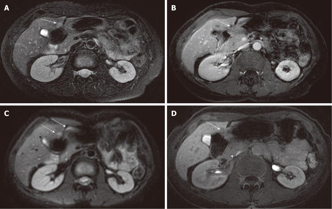
Figure 4 Metastasis in the IV segment.
A: A metastatic lesion in colon carcinoma is depicted in the most caudal area of the IV liver segment, it was well depicted in the fat-suppressed fast spin-echo T2-weighted sequence (arrow); B: In the gadolinium-enhanced acquisition (arrow); C: The lesion is poorly represented in the diffusion-weighted imaging, covered by magnetic susceptibility artifacts due to the adjacent intestinal loop (arrows); D: Delayed imaging after gadolinium-ethoxybenzyl-diethylenetriamine pentaacetic acid confirmed the metastatic lesion (arrow).
- Citation: Palmucci S, Mauro LA, Messina M, Russo B, Failla G, Milone P, Berretta M, Ettorre GC. Diffusion-weighted MRI in a liver protocol: Its role in focal lesion detection. World J Radiol 2012; 4(7): 302-310
- URL: https://www.wjgnet.com/1949-8470/full/v4/i7/302.htm
- DOI: https://dx.doi.org/10.4329/wjr.v4.i7.302


