Copyright
©2012 Baishideng Publishing Group Co.
World J Radiol. Jun 28, 2012; 4(6): 283-285
Published online Jun 28, 2012. doi: 10.4329/wjr.v4.i6.283
Published online Jun 28, 2012. doi: 10.4329/wjr.v4.i6.283
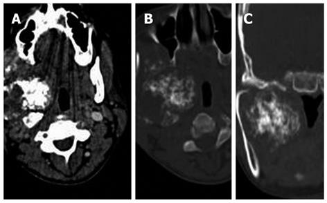
Figure 4 Contrast-enhanced computed tomography of the face and neck.
A: Axial contrast-enhanced computed tomography (CECT) section in soft tissue settings at the level of the maxillary antra reveal a lobulated soft tissue mass in the right parapharyngeal space with abundant chondroid matrix; B: An axial CECT bone window setting shows no destruction of the mandible; C: Coronal reformatted section in the bone window setting shows the pressure effect of the mass over the right mandible causing scalloping of the inner cortex of the condyle and the body.
- Citation: Gupta P, Bhalla AS, Karthikeyan V, Bhutia O. Two rare cases of craniofacial chondrosarcoma. World J Radiol 2012; 4(6): 283-285
- URL: https://www.wjgnet.com/1949-8470/full/v4/i6/283.htm
- DOI: https://dx.doi.org/10.4329/wjr.v4.i6.283


