Copyright
©2012 Baishideng Publishing Group Co.
World J Radiol. Jun 28, 2012; 4(6): 283-285
Published online Jun 28, 2012. doi: 10.4329/wjr.v4.i6.283
Published online Jun 28, 2012. doi: 10.4329/wjr.v4.i6.283
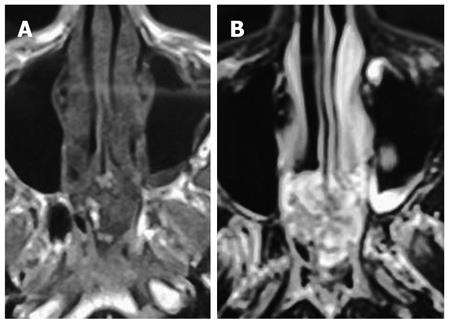
Figure 2 Magnetic resonance imaging of the skull base in the same patient.
A: Axial T1-weighted magnetic resonance imaging (MRI) demonstrates the hypointense mass arising from the sphenoid sinus; B: On axial T2-weighted MRI, the mass is predominantly hyperintense with a few hypodense areas.
- Citation: Gupta P, Bhalla AS, Karthikeyan V, Bhutia O. Two rare cases of craniofacial chondrosarcoma. World J Radiol 2012; 4(6): 283-285
- URL: https://www.wjgnet.com/1949-8470/full/v4/i6/283.htm
- DOI: https://dx.doi.org/10.4329/wjr.v4.i6.283


