Copyright
©2012 Baishideng Publishing Group Co.
World J Radiol. Jun 28, 2012; 4(6): 283-285
Published online Jun 28, 2012. doi: 10.4329/wjr.v4.i6.283
Published online Jun 28, 2012. doi: 10.4329/wjr.v4.i6.283
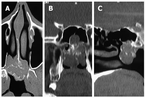
Figure 1 Non-contrast computed tomography of the paranasal sinuses.
A: Axial non-contrast computed tomography shows a soft tissue mass of the sphenoid sinus causing blockade of the posterior choanae. There is pleomorphic calcification in the center as well as the periphery of the mass not conforming to a particular type of matrix mineralization; B: Coronal reformation shows the origin of the mass from the floor of the sphenoid sinus; C: Sagittal reformation also demonstrates the mass arising from the floor of the sphenoid sinus.
- Citation: Gupta P, Bhalla AS, Karthikeyan V, Bhutia O. Two rare cases of craniofacial chondrosarcoma. World J Radiol 2012; 4(6): 283-285
- URL: https://www.wjgnet.com/1949-8470/full/v4/i6/283.htm
- DOI: https://dx.doi.org/10.4329/wjr.v4.i6.283


